Southern Illinois University
HISTO HOME
ERG Index
GI Index

ENDO Index

REPRO Index
RESOURCE CENTER

Hints for Slide ERG 5.  

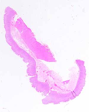
The source for this specimen should be obvious (and confidently so), provided you are familiar with the organ it represents.

Note the several distinct layers.

  • Name these layers.

Which organs have such distinct layers?

Note specific features of the mucosal layer.

  • What shape is the surface?
  • How many different kinds of epithelial cells comprise the surface epithelium?
  • Is lamina propria conspicuous?
  • Are there any epithelial features (e.g., glands or crypts) embedded in the lamina propria?
  • Can you find a muscularis mucosae?

Are there any distinctive features in the submucosa?

Is the muscularis externa organized into distinct layers?

Hints on the next page are a bit more pointed.  Don't look unless you are stuck.

More hints.  


Comments and questions: dgking@siu.edu

SIUC / School of Medicine / Anatomy / David King

https://histology.siu.edu/erg/SAQ/ERG05a.htm
Last updated:  4 September 2021 / dgk