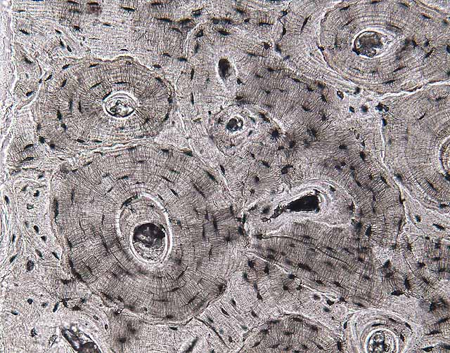


This image illustrates compact or cortical bone, sectioned across the long axis of the bone.
The specimen in this slide is "ground bone," a sliver of bone from which all organic materials (cells and collagen) have been removed, so we see here only the remaining mineral structure. Bone dust, a byproduct of the grinding process by which the specimen was prepared, can be found in Haversian canals.The most prominent features are the sets of concentric lamellae, called osteons, each with a Haversian canal at its center.
In life, each Haversian canal contains a blood vessel which brings nutrients to the tissue. Osteocytes occupy lacunae (here visible as small dark oblongs interspersed among the lamellae) interconnected by canaliculi.
Historical note: The eponym "Haversian" commemorates Clopton Havers, b. 1657.
Click on a thumbnail below for additional information.
Comments and questions: dgking@siu.edu
SIUC / School
of Medicine / Anatomy / David
King
https://histology.siu.edu/ssb/NM035b.htm
Last updated: 15 November 2021 / dgk