SIU labs can bring the THUNDER with new 3D imaging microscope
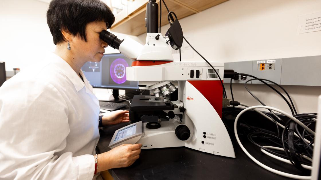
A new microscope at SIU School of Medicine can quickly make the invisible visible. The THUNDER ImagerTM, a highly advanced technology now in use at SIU’s Core Research Facilities, lets scientists capture vivid 3D images of cells, tissues and organs in seconds. The new capability opens the door to faster medical discoveries and groundbreaking visuals.
Scientists say the THUNDER ImagerTM could accelerate progress in areas like cancer, hearing loss and Alzheimer’s disease by helping scientists understand the body at the molecular level. SIU’s facility is now one of the few in downstate Illinois with this advanced imaging technology.
The advanced microscope developed by Leica Microsystems, employs technology to reduce out-of-focus light in fluorescence microscopy of thick specimens. This method enables high-resolution, high-speed imaging of various samples, including tissue sections and 3D cell cultures.
"The THUNDER Imager is a big step forward for our team,” said Melissa Roberts, assistant director of the SIU core laboratory. “It gives our scientists the ability to see complex structures in real time—and in remarkable detail. That kind of clarity can make a real difference in advancing our research.”
Our new innovative microscope can observe cellular interactions within organs and produce high-quality visuals for scientists in a fraction of the time previously required. This increased efficiency enables scientists to conduct more extensive studies and analyze larger datasets, potentially leading to more robust research outcomes.
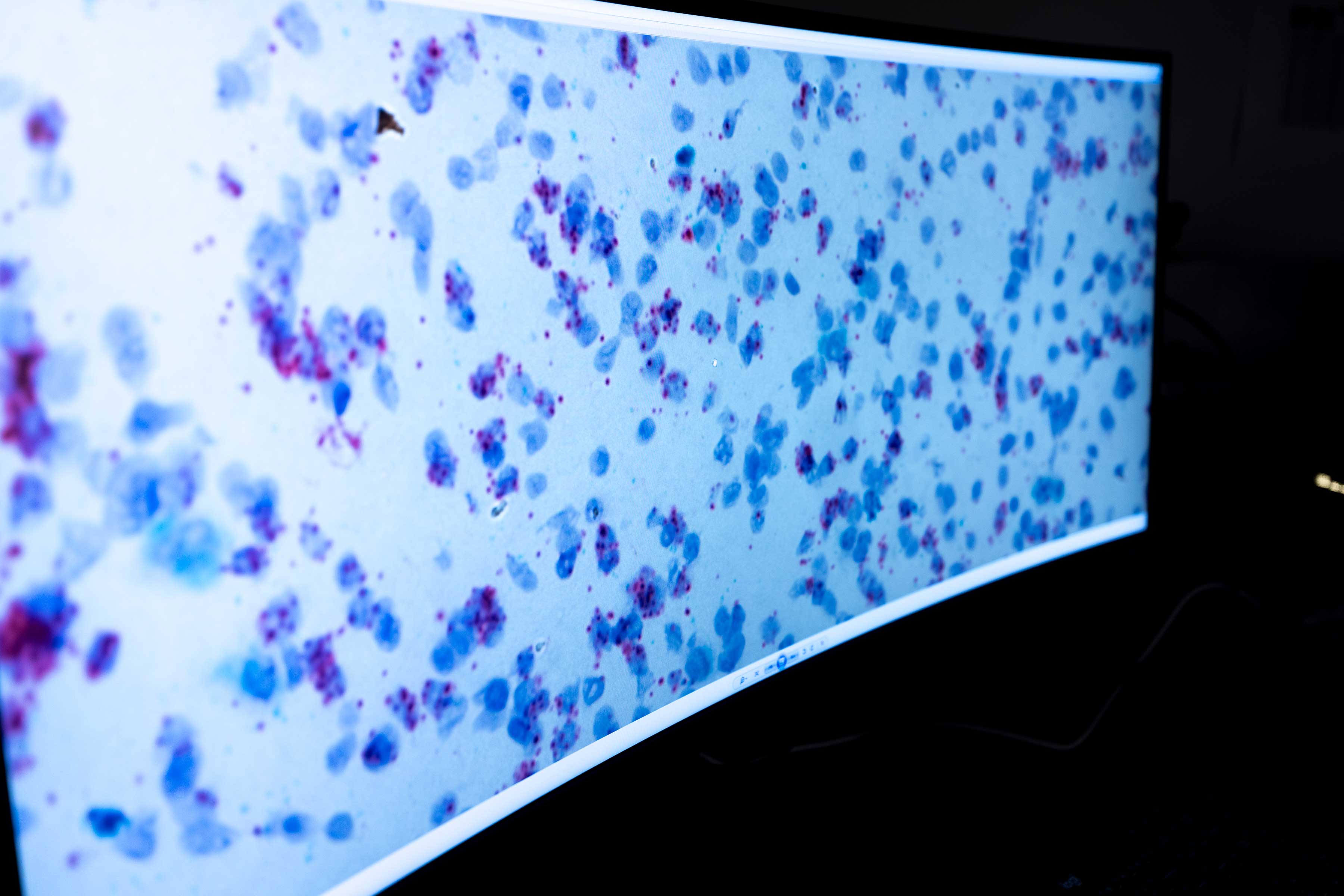 The system also supports a broad range of research applications, including live cell imaging, cleared tissue analysis and the investigation of complex 3D cultures. Its versatility provides new perspectives for exploring biological questions at multiple scales.
Donald Caspary, PhD, a pharmacology scientist at SIU School of Medicine, is using the microscope in his studies of age-related hearing loss. His work focuses on changes in brain receptors involved in hearing. The THUNDER ImagerTM allows his team to scan brain tissue much faster and capture higher-quality images, allowing more accurate measurements to save time and improve results.
SIU School of Medicine Core Research Facilities provides investigators with access to leading-edge technologies. The acquisition of the THUNDER ImagerTM reflects this commitment and strengthens the institution's position in biomedical research.